Craniotomy vs Craniectomy: Understanding the Key Differences
When facing neurological conditions requiring surgical intervention, understanding the nuances between procedures is crucial. Two common procedures involving the skull are craniotomy and craniectomy. While both involve accessing the brain, they differ significantly in their execution and purpose. This article delves into the key differences between a craniotomy and a craniectomy, providing a comprehensive understanding of each procedure, their indications, risks, and recovery processes. Choosing the right procedure is vital; it impacts patient outcomes significantly. Let’s explore these differences in detail.
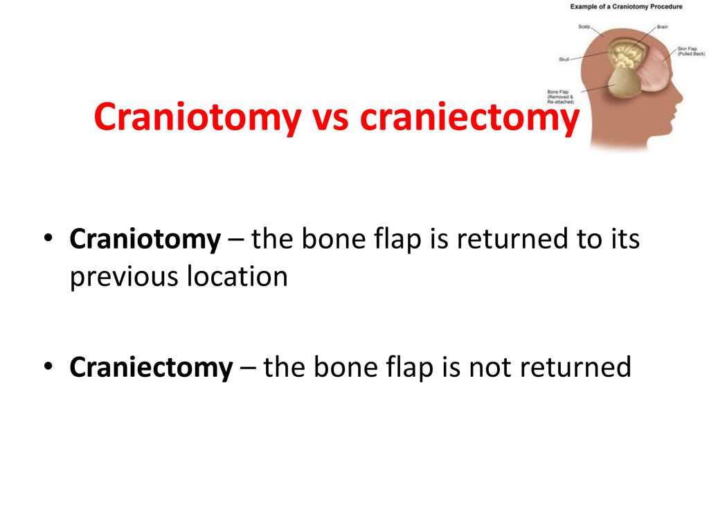
What is a Craniotomy?
A craniotomy is a surgical procedure where a section of the skull, called a bone flap, is temporarily removed to access the brain. After the necessary procedure on the brain is completed, the bone flap is typically replaced and secured back into its original position using plates, screws, or sutures. This allows the skull to heal naturally, providing protection to the brain. A craniotomy is often performed for a variety of reasons, including:
- Removing brain tumors
- Repairing aneurysms
- Evacuating hematomas (blood clots)
- Treating arteriovenous malformations (AVMs)
- Relieving pressure inside the skull
The primary goal of a craniotomy is to gain access to the brain to treat a specific condition while preserving the structural integrity of the skull. Precise planning and execution are paramount to minimize risks and maximize the benefits of the surgery.
The Craniotomy Procedure
The craniotomy procedure generally involves the following steps:
- Preparation: The patient is placed under general anesthesia. The scalp is shaved and cleaned with an antiseptic solution.
- Incision: A surgical incision is made in the scalp. The size and location of the incision depend on the area of the brain that needs to be accessed.
- Bone Flap Creation: The surgeon uses a specialized drill to create a circular or rectangular bone flap. This flap is carefully lifted and removed, exposing the dura mater (the protective membrane surrounding the brain).
- Brain Access: The dura mater is opened to expose the brain. The surgeon then performs the necessary procedure, such as tumor removal or aneurysm repair.
- Closure: After the procedure is complete, the dura mater is closed, and the bone flap is carefully replaced and secured. The scalp incision is then closed with sutures or staples.
What is a Craniectomy?
A craniectomy is a surgical procedure similar to a craniotomy, but with a crucial difference: the bone flap that is removed is not immediately replaced. Instead, the bone flap is typically stored (either cryopreserved or implanted in the patient’s abdomen) or discarded. The opening in the skull is left to allow the brain to swell without being compressed. This is particularly important in cases where the brain is likely to swell significantly due to trauma, stroke, or other conditions. A craniectomy is commonly performed in the following situations:
- Severe traumatic brain injury (TBI)
- Stroke with significant swelling
- Malignant cerebral edema
- Decompressive surgery to reduce intracranial pressure
The primary goal of a craniectomy is to relieve pressure inside the skull and prevent further brain damage caused by swelling. Leaving the bone flap out allows the brain to expand and reduces the risk of compression.
The Craniectomy Procedure
The craniectomy procedure shares many similarities with the craniotomy procedure, but with the key difference of leaving the bone flap out. The general steps include:
- Preparation: The patient is placed under general anesthesia. The scalp is shaved and cleaned.
- Incision: A surgical incision is made in the scalp.
- Bone Flap Removal: A bone flap is created using a drill and carefully removed.
- Brain Access: The dura mater is opened to expose the brain. The surgeon performs the necessary procedure.
- Closure: The dura mater is closed, but the bone flap is not replaced. The scalp incision is closed, leaving the opening in the skull covered only by the scalp and underlying tissues.
Key Differences: Craniotomy vs Craniectomy
The most significant difference between a craniotomy and a craniectomy lies in whether the bone flap is replaced after the surgical procedure. Here’s a summary of the key distinctions:
- Bone Flap Replacement: In a craniotomy, the bone flap is replaced. In a craniectomy, it is not.
- Purpose: A craniotomy is typically performed to access and treat specific brain conditions while maintaining the skull’s integrity. A craniectomy is primarily performed to relieve pressure inside the skull due to brain swelling.
- Indications: Craniotomies are used for tumor removal, aneurysm repair, and other procedures where brain swelling is not a major concern. Craniectomies are used for severe TBI, stroke with swelling, and other conditions where relieving pressure is critical.
- Recovery: Recovery after a craniectomy can be more complex due to the absence of the bone flap. A subsequent procedure, called a cranioplasty, is often required to replace the bone flap at a later date.
When is Each Procedure Necessary?
The decision to perform a craniotomy or a craniectomy depends on the specific clinical situation and the patient’s condition. Here’s a more detailed look at when each procedure is typically indicated:
Craniotomy Indications
- Brain Tumors: Craniotomy is the standard approach for removing brain tumors. It allows surgeons to access the tumor while preserving the surrounding brain tissue.
- Aneurysms: Repairing brain aneurysms often requires a craniotomy to clip or coil the aneurysm, preventing it from rupturing.
- Arteriovenous Malformations (AVMs): Craniotomy is used to surgically remove AVMs, which are abnormal tangles of blood vessels in the brain.
- Hematomas: Evacuating blood clots (hematomas) from the brain may require a craniotomy to access and remove the clot.
Craniectomy Indications
- Traumatic Brain Injury (TBI): In cases of severe TBI with significant brain swelling, a craniectomy can be life-saving. Removing the bone flap allows the brain to swell without being compressed, reducing the risk of further damage.
- Stroke: A craniectomy may be necessary after a large stroke to relieve pressure caused by swelling in the affected area of the brain.
- Malignant Cerebral Edema: Conditions causing severe brain swelling, such as malignant cerebral edema, may require a craniectomy to prevent herniation and death.
Risks and Complications
Both craniotomy and craniectomy are complex surgical procedures that carry potential risks and complications. These can include:
- Infection: Infection at the surgical site or within the brain is a risk with any surgical procedure.
- Bleeding: Bleeding during or after surgery can lead to hematoma formation and increased pressure inside the skull.
- Blood Clots: Blood clots can form in the legs or lungs and travel to the brain, causing a stroke.
- Seizures: Seizures can occur after either procedure, particularly if the brain tissue is irritated or damaged.
- Neurological Deficits: Damage to brain tissue during surgery can lead to neurological deficits such as weakness, speech problems, or cognitive impairment.
- Cerebrospinal Fluid (CSF) Leak: CSF can leak from the surgical site, requiring additional treatment.
- Hydrocephalus: Hydrocephalus (accumulation of fluid in the brain) can occur after surgery, requiring a shunt to drain the fluid.
- Sinkin Skin Flap Syndrome: After a craniectomy, where the bone flap is removed, the scalp may sink inwards due to atmospheric pressure.
The specific risks and complications can vary depending on the patient’s condition, the location of the surgery, and the surgeon’s experience. Patients should discuss these risks with their surgeon before undergoing either procedure. [See also: Brain Surgery Recovery Tips]
Recovery and Rehabilitation
Recovery after a craniotomy or craniectomy can be a lengthy process, and the specific recovery plan will depend on the individual patient’s condition and the extent of the surgery. Generally, the recovery process involves:
- Hospital Stay: Patients typically stay in the hospital for several days to weeks after surgery.
- Pain Management: Pain medication is used to manage pain and discomfort after surgery.
- Physical Therapy: Physical therapy helps patients regain strength, mobility, and coordination.
- Occupational Therapy: Occupational therapy helps patients regain skills needed for daily living.
- Speech Therapy: Speech therapy helps patients regain speech and language skills if they have been affected by the surgery.
- Cognitive Rehabilitation: Cognitive rehabilitation helps patients improve their memory, attention, and problem-solving skills.
- Cranioplasty (for Craniectomy): After a craniectomy, a cranioplasty procedure is typically performed to replace the bone flap. This is usually done several months after the initial surgery, once the brain swelling has subsided.
The rehabilitation process can be challenging, but with dedication and support, patients can often make significant progress in their recovery. [See also: Understanding Brain Swelling After Surgery]
Cranioplasty: Reconstructing the Skull After Craniectomy
As mentioned, a cranioplasty is a surgical procedure performed to repair a skull defect created by a craniectomy. The bone flap removed during the craniectomy is either replaced, or a synthetic material is used to reconstruct the missing portion of the skull. The timing of the cranioplasty depends on several factors, including the patient’s overall health, the presence of infection, and the degree of brain swelling. Ideally, the cranioplasty is performed several months after the craniectomy, allowing the brain to fully recover and the swelling to subside. The main benefits of cranioplasty include:
- Protection of the Brain: The reconstructed skull provides protection to the underlying brain tissue.
- Improved Cosmesis: Cranioplasty can improve the appearance of the skull, reducing the sunken appearance that can occur after a craniectomy.
- Neurological Improvement: Some patients experience improvements in neurological function after cranioplasty, such as improved cognitive function or motor skills.
Conclusion
Craniotomy and craniectomy are distinct surgical procedures with different purposes and indications. A craniotomy involves temporarily removing and replacing a bone flap to access the brain for various treatments, while a craniectomy involves removing a bone flap and not immediately replacing it, primarily to relieve pressure caused by brain swelling. Understanding the differences between these procedures is essential for patients and their families facing neurological conditions requiring surgical intervention. The choice between a craniotomy and a craniectomy depends on the specific clinical situation and the patient’s individual needs. Both procedures carry risks and require a comprehensive recovery and rehabilitation plan. By working closely with their medical team, patients can make informed decisions and optimize their outcomes. Ultimately, whether a patient undergoes a craniotomy or craniectomy, the goal remains the same: to improve their neurological health and quality of life. Furthermore, a follow-up cranioplasty after a craniectomy can offer additional benefits, including protection, cosmetic improvement, and potentially enhanced neurological function.
